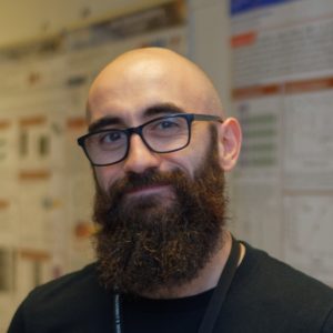
eric.clot
On Wednesday April 17th, at 14:00 Eric Clot (SPINTEC) will defend in English his PhD thesis entitled :
Nuts and bolts approach to developing a nitrogen-vacancy microscope inside an electromagnet
Place : IRIG/SPINTEC, CEA Building 10.05, auditorium 445 (access needs authorization request it before 7th to admin.spintec@cea.fr)
video conference : https://cnrs.zoom.us/j/92705963806?pwd=V0xDbkFsUXJiNE0wanI3SEYrK2lxQT09
Meeting ID: 927 0596 3806 Passcode: Eu1igm
Abstract : The ability to optically detect the electron paramagnetic resonance (EPR) of a single nitrogen-vacancy (NV) defect in a single diamond crystal has transformed the field of spectroscopy. By attaching a nano-diamond containing a single color defect to the tip of a near-field microscope cantilever, one could build a scanning spectrometer that could locally map the modulation of its EPR response without compromising frequency range, external magnetic field strength, spatial resolution, non-perturbation of the equilibrium texture, sensitivity, or accuracy. However, the general use of such an instrument would require the ability to apply an external magnetic field. This is still an enormous technological challenge, since it requires a solution that allows high-precision control of both the amplitude and the orientation of the external magnetic field. The aim of my thesis was to take up this challenge by focusing on the development of an NV center microscope that fits between the poles of an ultra-stable and orientable electromagnet. This initiative is a collaboration between Spintec and the Institut Néel. This approach introduced significant space constraints on the optical block and the optical interferometer. To this end, I developed a novel type of ultra-compact high performance optical objectives with large numerical aperture, high optical transmission and zero achromaticity. The design is based on a purely achromatic reflective design composed of four axisymmetric surfaces, including two conic elements – a parabola and an ellipse – that share a focal point. This invention is patented. At the same time, an optical bench NV microscopy setup was established to accelerate the development of measurement protocols free from the spatial constraints of using an electromagnet. This setup allows direct evaluation of NV centers and samples by integrating a high performance confocal optical circuit, microwave instrumentation, precision actuators, and timing accurate devices. This apparatus allowed the formulation and implementation of protocols to characterize NV centers in AFM tips, precisely analyzing their properties such as orientations or Rabi frequencies. In addition, it facilitated the first magnetometric assessments on samples using the fluorescence quenching method, validating the system’s ability to generate stray magnetic field maps. The next goals of the project include the refinement of the pulsed sequence protocol to image the spatio-temporal profile of standing spin waves in confined geometries. These methods will first be applied to the optical table NV microscope, with the aim of rapidly extending their functionalities to the microscope operating in the electromagnet.
titre : «Mise au point d’un microscope à centre azote-lacune dans un électro-aimant»
résumé : La capacité de détecter optiquement la résonance paramagnétique électronique (RPE) d’un défaut unique lacune-azote dans un cristal de diamant (appelé aussi centre NV dans sa dénomination anglaise pour ‘nitrogen-vacancy’) a transformé le domaine de la spectroscopie. En attachant un nano-diamant contenant un tel défaut coloré unique à la pointe d’un cantilever de microscope à champ proche, on pourrait construire un spectromètre à balayage capable de cartographier localement la modulation de sa réponse RPE sans compromettre la gamme de fréquences, l’intensité du champ magnétique externe, la résolution spatiale, l’absence de perturbation de la texture d’équilibre, la sensibilité ou la précision. Cependant, l’utilisation générale d’un tel instrument nécessiterait la capacité d’appliquer un champ magnétique externe. Il s’agit encore d’un énorme défi technologique, puisqu’il nécessite une solution permettant un contrôle de haute précision de l’amplitude et de l’orientation de ce champ magnétique. L’objectif de ma thèse était de relever ce défi en se concentrant sur le développement d’un microscope à centre NV qui s’insère entre les pôles d’un électro-aimant ultra-stable et orientable. Cette initiative est une collaboration entre Spintec et l’Institut Néel. Cette approche a introduit des contraintes d’espace significatives sur le bloc optique et l’interféromètre optique. À cette fin, j’ai développé un nouveau type d’objectifs optiques ultra-compacts à haute performance avec une grande ouverture numérique, une transmission optique élevée et une achromaticité nulle. La conception est basée sur un motif catoptrique intrinsèquement achromatique composé de quatre surfaces axisymétriques, dont deux éléments coniques – une parabole et une ellipse – qui partagent un point focal. Cette invention est brevetée. Parallèlement, un banc optique de microscopie NV a été mis en place pour accélérer le développement de protocoles de mesure libérés des contraintes spatiales liées à l’utilisation d’un électro-aimant. Cette installation permet l’évaluation directe des centres NV et des échantillons en intégrant un circuit optique confocal à haute performance, une instrumentation à micro-ondes, des actionneurs de précision et des dispositifs de synchronisation précis. Cet appareil a permis de formuler et de mettre en œuvre des protocoles pour caractériser les centres NV dans les pointes AFM, en analysant précisément leurs propriétés telles que les orientations ou les fréquences de Rabi. En outre, il a facilité les premières évaluations magnétométriques sur des échantillons en utilisant la méthode du « quenching » de la fluorescence, validant la capacité du système à générer des cartes de champs de fuites magnétiques. Les prochains objectifs du projet comprennent l’affinement du protocole de séquence pulsée afin d’obtenir des images du profil spatio-temporel des ondes de spin stationnaires dans des géométries confinées. Ces méthodes seront d’abord appliquées au microscope NV à table optique, dans le but d’étendre rapidement leurs fonctionnalités au microscope fonctionnant dans l’électro-aimant.
Jury :
- Claude Fermon, Directeur de recherche, SPEC – Univ. Paris-Saclay CNRS CEA, rapporteur
- Daniel Lacour, Directeur de recherche, Institut Jean Lamour, rapporteur
- Christian Degen, Professeur, ETH Zürich, Examinateur
- Aurore Finco, Chargée de recherche, Laboratoire Charles Coulomb, Examinatrice
- Hélène Béa, Chargée de recherche HDR, SPINTEC, Examinatrice
Encadrants :
- Olivier Klein, Directeur de recherche, Spintec, CEA Grenoble, Directeur de thèse
- Benjamin Pigeau, Chargé de recherche, Institut Néel, Co-encadrant
Invités :
- Isabelle Joumard, Ingénieure, SPINTEC


